

Schedule an Exam Today
About Eye Tech Eyes
Established in 1993, The Eye Doctors at Eye Tech Eye Associates are experienced optometrists who pride themselves in staying up to date on the latest advanced technology and procedures in their field. If you live in Glendale, Peoria, Phoenix or Sun City call us to set up an appointment and meet one of our talented ODs.


Meet our Eye Doctors


Patient Reviews
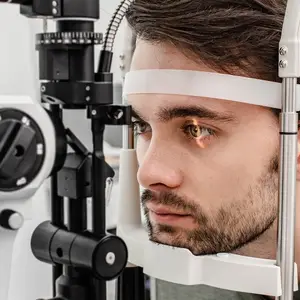
Our Eye Care Services at Eye Tech Eyes in Peoria, Arizona

Dry Eyes
Do you have dry, itchy, gritty-feeling eyes? Dry eye syndrome is a very common condition. We offer dry eye treatments at our eye care clinic.

Comprehensive Eye Exams
From updating your eyeglasses prescription to detection and treatment of eye diseases, comprehensive eye exams are important for continued visual health.
Contact Lens Exams
During a contact lens exam, your eye doctor will check if you are a good candidate for contacts and find the best type of contacts for your needs.
Eyeglasses & Frames

Designer Frames
Our extensive optical section offers a wide variety of eyeglass frames in every style, material & design. Come visit us today to see for yourself!
Our expert optical team can find just the right pair of glasses for you to be confident and look your best.
Lens coatings improve visual comfort, make it easier to clean your glasses and ensure your lenses last longer. Coatings include anti-scratch, anti-reflective, photochromatic and UV / blue light filters.
Contact Lenses

Contact Lens Fitting
We offer a wide range of contact lens options from dailies, monthly to multifocal contact lenses for crystal clear vision and superior comfort.
During a contact lens exam, your eye doctor will check if you are a good candidate for contacts and find the best type of contacts for your needs.
Health & Wellness

EyePromise Vitamins®
The #1 doctor-recommended vitamin company with the most innovative, highest-quality eye vitamins available.

MacuHealth Supplements®
MacuHealth is a leader in the eye supplement industry that is focused on innovation. Providing premium products formulated with pure, stable ingredients, MacuHealth cares for the eye at every stage of life.

Buy One Get One 50% Off
Q2 Promo 2026 BIGGEST SAVINGS OF THE SEASON (Apr 6 - Jun 14): Buy One Get One 50% Off!
*Must present offer at time of purchase. Must purchase a complete pair of prescription eyeglasses, including frame and lenses. Discount applied to complete pair of equal or lesser value. Does not include Barton Perreira, Burberry, Cartier, Cazal, Chanel, Cutler and Gross, Dita Lancier, Gucci, ic!Berlin, l.a. Eyeworks, Maui Jim, Mykita, Nifties, Oakley, Oliver Peoples, Ray-Ban, Salt, Silhouette, Tom Ford, WOOW, YSL, sunglass frames, accessories, contact lenses, or medical procedures. Cannot be used with any other promotion, discount, or offer. Not redeemable for cash. Not valid on previous orders. Other restrictions may apply. Offer ends 06/14/2026.

Save $400 on ACUVUE
We Accept Most Insurance Plans
Have another plan?
Benefit questions?
Paying out of pocket?
Call us, we are happy to help.


Location & Opening Hours
18431 N 91st Ave #1, Peoria, AZ 85382
- Onsite Parking
- Wheelchair Accessible
- Shop Glasses
No appointment needed
- Near Public Transit


- Fri Jul. 3 Closed
- Sat Jul. 4 Closed
- Mon Sep. 7 Closed
- Thu Nov. 26 Closed
- Fri Nov. 27 Closed
- Fri Dec. 25 Closed
- Monday 8:00 am - 4:30 pm
- Tuesday 8:00 am - 4:30 pm
- Wednesday 8:00 am - 4:30 pm
- Thursday 8:00 am - 4:30 pm
- Friday 7:00 am - 12:00 pm
- Saturday Closed
- Sunday Closed
We are excited to share ACUVUE® VITA® for Astigmatism will offer expanded around‑the‑clock parameters up to -2.75 cylinder, beginning March 23, 2026, giving broader flexibility to meet more patients’ needs. With this enhancement, ACUVUE®'s full reusable portfolio—including ACUVUE® OASYS 2‑Week for Astigmatism—will now provide coverage for up to 96% of astigmatic patients. ACUVUE® VITA® product family is a smart choice for new wearers— delivering superior comfort and vision all month long at an amazing value. Plus, keep an eye out for ACUVUE® VITA®'s fresh new look with updated packaging coming soon.